Automated Breast Ultrasound
Automated Breast Ultrasound
Enhanced Imaging for Dense Breast Tissue
Breast cancer screening using ultrasound helps doctors find cancers in dense breast tissue, which can be missed by traditional mammography. If you have dense breast tissue, using automated breast ultrasound (ABUS) screening with mammography has demonstrated a 35.7% increase in cancer detection compared to mammography alone.
ABUS uses sound waves that we use to create 3D images, allowing our radiologists to view hundreds of breast tissue “slices” that can show cancer or lesions in dense breast tissue that traditional imaging techniques might miss.
What to Expect
ABUS screening isn’t like a mammogram. We apply a layer of lotion and then place a scanner on your breast to begin taking images. The exam usually takes about 15 minutes and produces clear 3D ultrasound images. Your doctor then reviews the ABUS images alongside your mammogram.
Why Breast Density Matters
Why Breast Density Matters
Breasts are made of fat and breast tissue. A breast with more tissue than fat is considered dense. Breast density is determined by the radiologist who reads your mammogram. There are four density categories: A, B, C, and D. C and D are considered dense.1 Ask your doctor your density; every woman should know her breast density.
On a mammogram, dense tissue and masses both appear white, so a suspicious lump may be hidden in dense tissue. When dense tissue is scanned with ultrasound, tissue appears white and masses appear black – making them easier to see. Having dense breast tissue may also increase the risk of developing breast cancer 4 to 6 times.2
- Breast Imaging and Reporting and Data System (BI-RADS®), American College of Radiology.
- Boyd et al, New England Journal of Medicine 2007;356:227-36.
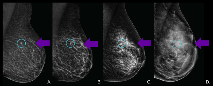
A. Almost entirely fatty. B. Scattered fibroglandular density. C. Heterogeneously dense. D. Extremely dense.
Hypothetical cancer in circle. This is easily seen in the breasts with fatty (A) and scattered (B) density, but it is obscured on the heterogenously (C) and extremely dense (D) breasts.